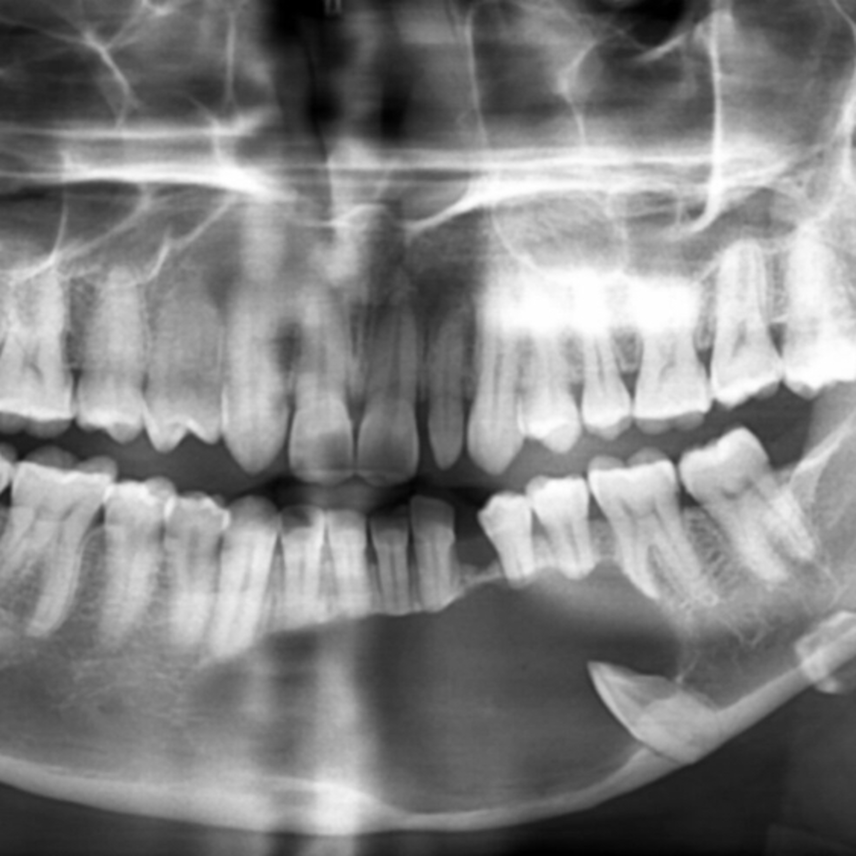
📝 "What Are Key Features Of The Adenomatoid Odontogenic Tumor?"
- Author
- Brendan Gallagher, DDS
- Published
- Sun 13 Apr 2025
- Episode Link
- https://podcasters.spotify.com/pod/show/doctorgallagher/episodes/What-Are-Key-Features-Of-The-Adenomatoid-Odontogenic-Tumor-e31fr88
Quick Review #276 - #pathology #oralpathology #doctorgallagher #oralsurgery #oralsurgeon #dentist #dentistry #dental #aot #adenomatoidodontogenictumor
- 4.13.25
Adenomatoid Odontogenic Tumor (AOT) is a benign (hamartomatous) epithelial odontogenic tumor most commonly affecting adolescents and young adults, with a strong predilection for females (2:1) and typically occurring during the second decade of life. It accounts for 3–7% of all odontogenic tumors.
Location:
- Maxillary anterior region is most frequently involved (up to 65% of cases).
- Often associated with an impacted tooth, especially the maxillary canine.
Clinical Presentation:
- Usually asymptomatic, slow-growing lesion.
- May present with painless swelling or delayed tooth eruption.
Radiographic Features:
- Appears as a well-circumscribed unilocular radiolucency.
- Frequently surrounds the crown and may extend apically beyond the CEJ, unlike a dentigerous cyst.
- Characteristic presence of fine, “snowflake”-like calcifications within the radiolucency in 70% of cases.
- Often causes mild expansion but rarely leads to root resorption.
Histopathology:
- Composed of spindle-shaped epithelial cells forming duct-like structures, rosettes, and whorled patterns.
- May contain eosinophilic amyloid-like material and dystrophic calcifications.
- The name “adenomatoid” reflects the duct-like structures microscopically, though no glandular differentiation is present.
Variants:
- Follicular type: associated with an impacted tooth.
- Extrafollicular type: not associated with a tooth but still intraosseous.
- Peripheral type: rare, presents in gingiva.
Treatment and Prognosis:
- Managed by simple enucleation.
- Excellent prognosis with rare recurrence due to its encapsulated nature.
Key Differentiators:
- More common in younger females, anterior maxilla, and often misdiagnosed as a dentigerous cyst or calcifying odontogenic cyst.
- Radiographic extension beyond the CEJ and presence of intra-lesional calcifications help distinguish it from other cystic lesions.
References:
1. Loganathan, K., & Vaithilingam, B. (2014). Adenomatoid odontogenic tumor in the mandible: A case report. International Journal of Dental Sciences and Research, 2(4B), 6–8.
2. Neville, B. W., Damm, D. D., Allen, C. M., & Chi, A. C. (2015). Oral and Maxillofacial Pathology (4th ed.). Saunders.
3. Philipsen, H. P., & Reichart, P. A. (1998). Adenomatoid odontogenic tumour: Facts and figures. Oral Oncology, 34(5), 389–399.
4. Buchner, A., Merrell, P. W., & Carpenter, W. M. (2006). Relative frequency of central odontogenic tumors: A study of 1,088 cases from Northern California and comparison to studies from other parts of the world. Journal of Oral and Maxillofacial Surgery, 64(9), 1343–1352.
5. ChatGPT. 2025.
#podcast #dentalpodcast #doctorgallagherpodcast #doctorgallagherspodcast #doctor #dentist #dentistry #oralsurgery #dental #dentalschool #dentalstudent #doctorlife #dentistlife #oralsurgeon #doctorgallagher
