📝 "What Are Key Characteristics Of The Juvenile Ossifying Fibroma?"
- Author
- Brendan Gallagher, DDS
- Published
- Tue 13 May 2025
- Episode Link
- https://podcasters.spotify.com/pod/show/doctorgallagher/episodes/What-Are-Key-Characteristics-Of-The-Juvenile-Ossifying-Fibroma-e32p9ut
Quick Review #278 - #pathology #oralpathology #doctorgallagher #oralsurgery #oralsurgeon #dentist #dentistry #dental
- 5.13.25
Location:
- Most commonly involves the maxilla, mandible, paranasal sinuses, or orbit.
- Maxilla is more frequently involved in the trabecular variant, while the psammomatoid variant commonly affects the orbit and paranasal sinuses.
Demographics:
- Occurs predominantly in children and adolescents (generally under age 15–20).
- No strong sex predilection, but some studies note a slight male predilection.
Radiographic Appearance:
- Well-circumscribed expansile radiolucent lesion, may show varying degrees of internal radiopacity due to mineralization.
- Cortical thinning and expansion are common; lesion may appear multilocular.
- The borders are typically well defined, sometimes with a sclerotic rim.
Histopathology:
2 main variants:
1 - Trabecular JOF: Characterized by trabeculae of immature woven bone in a cellular fibrous stroma.
2 - Psammomatoid JOF: Features small, round ossicles (psammoma-like bodies) in a fibrous stroma.
- Highly cellular, mitotic figures may be seen but no malignancy.
Variants:
- Trabecular Juvenile Ossifying Fibroma (TrJOF)
- Psammomatoid Juvenile Ossifying Fibroma (PsJOF)
Differential Diagnosis:
1. Fibrous dysplasia
2. Conventional ossifying fibroma
3. Aneurysmal bone cyst
4. Cemento-ossifying fibroma
5. Osteoblastoma
6. Low-grade osteosarcoma (due to aggressive growth in some cases)
Management:
A) Surgical excision or enucleation with curettage is the mainstay.
B) Requires long-term follow-up due to high recurrence rates (up to 30–50% in some cases).
C) In aggressive or recurrent cases, more extensive resection may be necessary.
References:
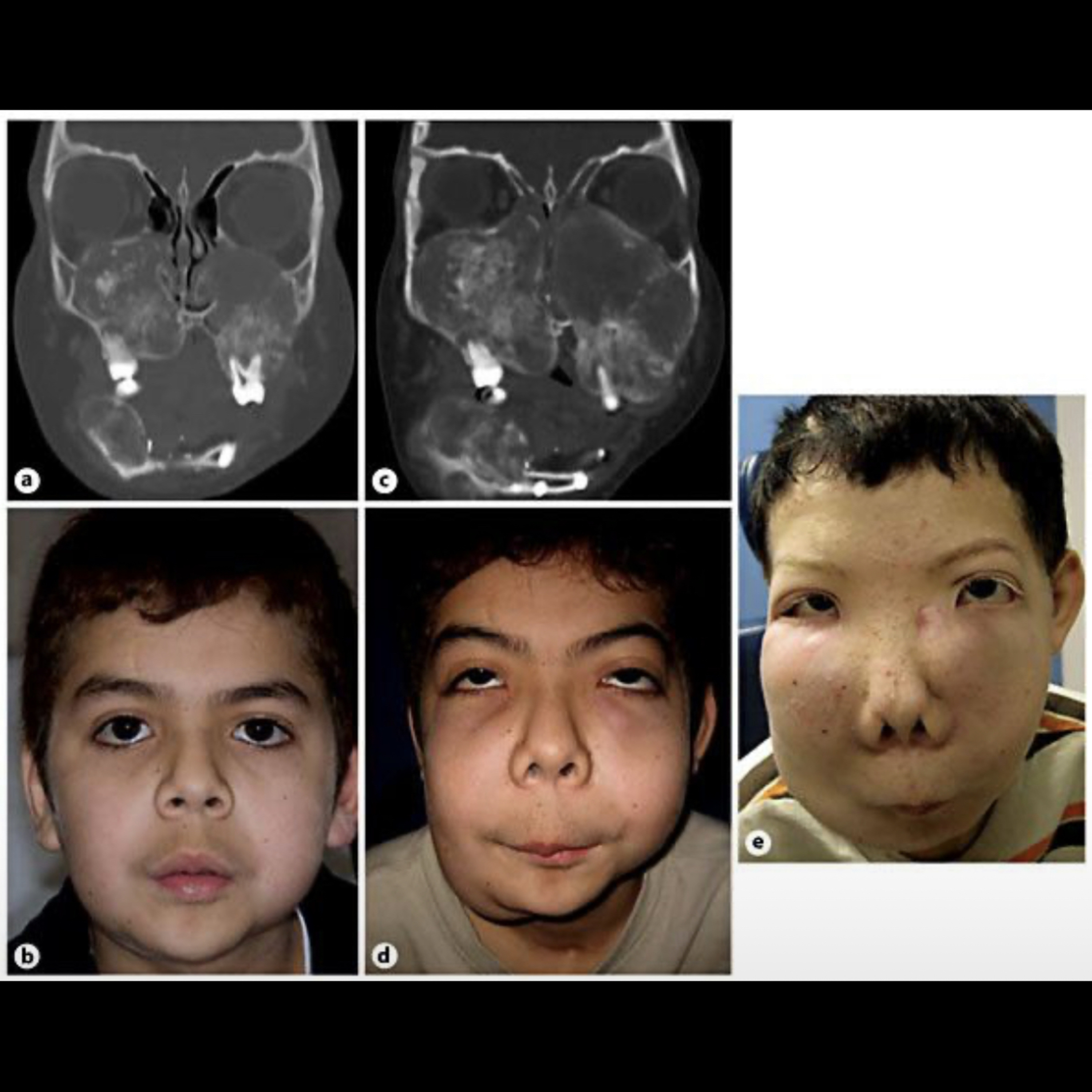
1. Merritt, H. A. (n.d.). Clinical findings in our patient with juvenile ossifying fibroma: a Coronal CT scan with contrast showing bilateral maxillary and right mandibular bone lesions [Figure]. ResearchGate.
2. Gautier, B., Dugast, S., Guyonvarc'h, P., Longis, J., Corre, P., & Bertin, H. (2024).
Ossifying fibroma and juvenile ossifying fibroma: A systematic review on clinical and radiological parameters, treatment modalities, and recurrence. Journal of Stomatology, Oral and Maxillofacial Surgery, 125(3), 102185.
3. Titinchi, F. (2021).
Juvenile ossifying fibroma of the maxillofacial region: A multi-center retrospective study. Journal of Oral and Maxillofacial Surgery, 79(8), 1672.e1–1672.e9.
4. ChatGPT.2025
#podcast #dentalpodcast #doctorgallagherpodcast #doctorgallagherspodcast #doctor #dentist #dentistry #oralsurgery #dental #dentalschool #dentalstudent #doctorlife #dentistlife #oralsurgeon #doctorgallagher